Abstract Lipids are important molecules for us for a number of reasons. These compounds are essential for living organisms as they are the major building blocks of cells, signal and energy storage molecules. Lipid metabolism and cellular signaling regulate cell growth, proliferation, and survival. They are one of the major constituents of foods, but over-consumption of certain lipid components can be detrimental to our health. Lipids are of great economic importance as agricultural products and they are major items of international commerce. Methods for the analysis of lipids are thus fundamental for research, clinical and quality control applications.This chapter shows some basic strategies for analyzing lipids using HPLC/MS, a powerful method that provides reliable qualitative and quantitative data.
LevelBasic
A huge number of existing chemically distinct structures makes the definition of ![]() lipids difficult.
lipids difficult.
From the point of view of analytical chemistry lipids represent a very complex mixture of compounds with various functional groups, chain lengths, double bond number and positions, exhibiting both structural isomerism and stereoisomerism. A total of lipids in a certain environment, e.g., cell or tissue is called a lipidome.
Examples of lipids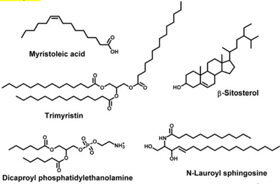 author
Lipid analysis usually begins with extracting all lipid constituents from the tissues. The extraction should be done as soon as possible after removal of the tissue from an organism, to avoid decomposition or alteration of lipids. A special pretreatment is usually carried out to deactivate enzymes and prevent autoxidation of polyunsaturated fatty acid chains.
- One of the most popular extraction methods uses chloroform-methanol (2:1), which forms ternary solvent system with water present in the sample. The method is referred as Folch extraction procedure and many modification of the original protocol can be found in literature.
- The Bligh and Dyer extraction method is recommended for large samples with high water content. Lipids are extracted with chloroform-methanol (1:2) and subsequently with pure chloroform.
- Plant tissues are recommended to extract first with 2-propanol in order to deactivate enzymes, particularly phospholipase D, which is set into action upon plant wounding.
In principle, two approaches are used for analysis of the total lipid extract:
- In the classical approach, lipid classes are separated from each other, usually by adsorption chromatography in TLC or column arrangement. Selected fractions (lipid classes) are then subjected to thorough analysis aiming at characterization of molecular species within the class.
- The second, lipidomic approach utilizes advanced mass spectrometric techniques to identify and quantify most of lipids at once, usually in a single run.
Lipids are usually separated using normal-phase, reversed-phase or silver ion chromatography. Unfortunately, neither separation system is able to fully resolve all components of the complex lipid mixtures, but each of them provides us with a valuable piece of information about the samples.
Normal-phase high-performance liquid chromatography (NP-HPLC)
In normal phase chromatography, the stationary ![]() phase is polar and the mobile phase is nonpolar. In NP-HPLC, the mobile phase is composed of organic solvents and the stationary phase is mostly silica or silica modified with polar groups, e.g., cyano or diol.
phase is polar and the mobile phase is nonpolar. In NP-HPLC, the mobile phase is composed of organic solvents and the stationary phase is mostly silica or silica modified with polar groups, e.g., cyano or diol.
- NP-HPLC is usually carried out using mobile phases without added water.
- A version of NP-HPLC that uses organic solvents (typically acetonitrile) with small amount of water is referred as hydrophilic interaction liquid chromatography (HILIC).
- NP-HPLC systems separate lipids based on polarity, i.e., according to the nature and number of polar functional groups (e.g. ester bonds, hydroxy or carboxy groups, glycosylation, etc.).
Separation of lipid standards by NP-HPLC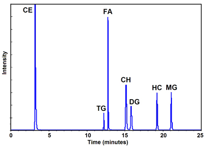 adapted from Genge et al., Anal. Biochem. 2003
Reversed-phase ![]() high-performance liquid chromatography (RP-HPLC)
high-performance liquid chromatography (RP-HPLC)
In RP-HPLC, the mobile phases are significantly more polar then the stationary phases, which are usually microporous silica chemically modified with octadecyl or octyl chains. Mobile phases consist of water or aqueous buffer solutions and various water-miscible solvents, e.g., methanol or acetonitrile. Separation in RP-HPLC is based on partitioning of the analytes between the hydrocarbon layer on the silica surface and the mobile phase. Selectivity is mostly determined by the mobile phase composition. Mobile phases for separating non-polar lipids are commonly prepared without water. Such systems are referred to as “non-aqueous reversed-phase” NARP.
Retention behaviour of neutral lipids (acylglycerols or fatty acid esters) is expressed in terms of Equivalent Carbon Number, ECN:
ECN = CN-2DB
Any double bond reduces retention in the same way as shortening the chain roughly by two carbon atoms. The elution order follows the ECN value, which is very useful for predicting retention behavior. Geometry of double bond(s) also affects retention; trans- isomers are usually retained more strongly than cis- counterparts.
Separation of natural mixture of triacylglycerols by NARP-HPLC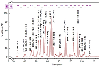 Author
Silver-ion high-performance liquid ![]() chromatography (Ag-HPLC)
chromatography (Ag-HPLC)
Ag-HPLC uses silver ions to form reversible charge-transfer complexes with π‑electrons of double bonds in the fatty acyl chains. Unsaturated analytes are donors of electrons and silver ions are their acceptors. Retention is a result of dynamic equilibria between the native and complexed analytes.
Retention times of lipids increases with the total number of double bonds in the fatty acid chains. Retention is also affected by double bond configuration and position within the chain; compounds with cis double bond geometry are bonded more strongly than the trans analogs. In polyenes, methylene-interrupted chains form more stable complexes than the conjugated ones and the strongest retention is observed for compounds with double bonds separated by two ![]() methylene groups. Compounds with a triple bond form weaker complexes than those with a double bond.
methylene groups. Compounds with a triple bond form weaker complexes than those with a double bond.
Separation of triacylglycerols by Ag-HPLC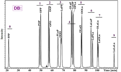 adapted from Lísa et al., Anal. Chem. 2009
Commercially available columns are based on ![]() cation-exchange phases, which minimize column bleeding. Less expensive alternative is a preparation of silver-ion column in the laboratory from a pre-packed cation-exchange column. Water is used as a mobile phase and silver nitrate solution is injected several times onto the column. Final step is replacement of water with organic solvents. Aprotic organic solvents, such as hexane, acetonitrile or acetone are preferred for Ag-HPLC.
cation-exchange phases, which minimize column bleeding. Less expensive alternative is a preparation of silver-ion column in the laboratory from a pre-packed cation-exchange column. Water is used as a mobile phase and silver nitrate solution is injected several times onto the column. Final step is replacement of water with organic solvents. Aprotic organic solvents, such as hexane, acetonitrile or acetone are preferred for Ag-HPLC.
Chiral-phase high-performance liquid chromatography
Stationary phases with chemically bonded chiral molecules are used for separation of enantiomeric lipids, particularly chiral diacylglycerols and monoacylglycerols. This approach avoids need for the preparation of diastereomeric derivatives.
Multidimensional separation systems
It is obvious that the best separation of complex lipid mixtures should be achieved by combination of the separation systems mentioned above. There is an increasing effort to develop reliable and efficient 2D chromatographic systems for lipids.
Most of the published work focuses on separation of triacylglycerol mixtures using combination of Ag-HPLC and NARP-HPLC. The analysis was originally performed off-line, when fractions from the first dimension (usually Ag-HPLC) are collected and re-analyzed in the second separation system (usually NARP-HPLC). The initial on-line setups worked in the stopped flow mode, but current comprehensive 2D systems operate continuously. Separation power of 2D systems is substantially enhanced over 1D approaches. The data are usually visualized as contour plots.
2D (Ag x NARP) separation of milk triacylglycerols 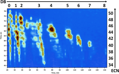 adapted from Dugo et al., J. Sep. Sci. 2006
Despite the current progress, on-line multidimensional separations are still not well established. Problems connected with incompatibility of mobile phases are encountered, which often lead to compromised separation conditions in both dimensions. Sensitivity is also usually reduced when compared with 1D systems. Nevertheless, research and instrumental development in this area promises interesting applications in the future.
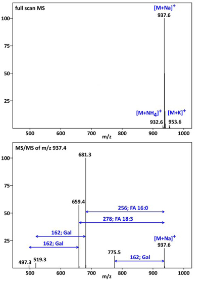
HPLC/MS combines the advantages of chromatography (high separation efficiency) and mass spectrometry (structural information, selectivity, sensitivity). Not surprisingly, MS detection gained increasingly wide usage also in the field of lipids. Almost all lipids can be easily ionized using one of the common atmospheric pressure ionization (API) techniques, i.e., by electrospray, atmospheric pressure chemical ionization or photoionization. The ions are analyzed directly or subjected to additional fragmentation(s) to get structural information. DGDG 18:3/16:0
Detection is highly selective and sensitive.
Electrospray ionization (ESI)
ESI is perhaps the most versatile API method. In ESI, the mobile phase enter the source through a capillary that has a high potential difference with respect to the sampling orifice. This forces the spraying of charged droplets. The solvent evaporates, droplets shrink and undergo multiple fissions, which finally results in charged analyte molecules.
ESI suits well for lipids with polar functional groups, e.g., phospholipids, glycolipids, free fatty acids, and sterols or oxidized lipids. It can also be applied for less polar or non-polar lipids, such are acylglycerols, sterol esters or wax esters. In the positive ion mode, lipids form either protonated molecules [M+H]+ or adducts with various alkali metals (Na+, K+, Li+), transition metal (Ag+) or other (e.g., NH4+) ions. Lipids with acidic group (e.g., phospholipids) are sensitively detected in the negative ion mode as deprotonated molecules [M-H]-.
Typically no fragments are found in ESI spectra. ESI is well compatible with water and water-miscible organic solvents (methanol, acetonitrile, etc.), but not with the non-polar ones, such as hexane.
ESI MS and MS/MS spectrum of digalactosyl diacylglycerol:
 author
![]() Nanoelectrospray ionization
Nanoelectrospray ionization
Nanoelectrospray is a miniaturized version of ESI suitable for spraying low amounts of low concentration samples. Disposable spray tips of a very small inner diameter are used. The ionization has an increased ![]() tolerance to highly aqueous solvents and salt contamination and very little sample clean up is being required.
tolerance to highly aqueous solvents and salt contamination and very little sample clean up is being required.
Atmospheric pressure ![]() chemical ionization (APCI)
chemical ionization (APCI)
In APCI, the mobile phase is heated to relatively high temperatures, (300-500°C), sprayed with high flow rates of nitrogen and the entire aerosol cloud is subjected to a corona discharge. The discharge forms reactant gas ions that undergo repeated collisions with the analyte, resulting in the formation of analyte ions. Unlike ESI, ionization in APCI is basically a gas phase process.
As APCI generates fragment ions, structural information can often be obtained even without MS/MS.
APCI MS spectrum of triacylglycerol TAG 18:1/26:2/18:1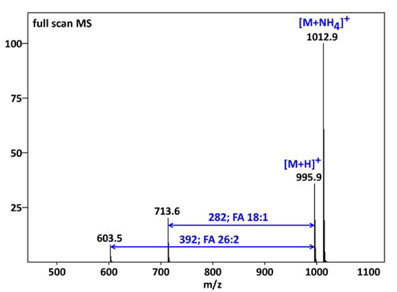 Author
Atmospheric pressure ![]() photoionization (APPI)
photoionization (APPI)
APPI is similar to APCI, but UV light accomplishes the ionization instead of corona discharge. In the direct APPI, the analyte molecule absorbs high-energy photons leading to ejection of one electron , resulting in a radical cation. The analyte radical cation can then react with the solvent by abstraction of a hydrogen to form stable [M+H]+, which is usually the observed ion. In dopant APPI, photoionizable compounds (e.g., toluene or acetone) are introduced into the ion source. The dopant ions are formed and react with neutral analyte molecules via proton transfer or charge exchange reactions.
When analyzing lipids by HPLC/MS, we can profit from ![]() fragmentation capabilities of the mass spectrometers. Fragmentation is usually achieved in the mass analyzer (MS/MS), but it can also take place in the ion source region of the spectrometer.
fragmentation capabilities of the mass spectrometers. Fragmentation is usually achieved in the mass analyzer (MS/MS), but it can also take place in the ion source region of the spectrometer.
Fragmentation spectra are very useful for elucidation of lipid structures. However, spectra interpretation is not always straightforward when two (or more) species co-elute. If the instrument allows only “in‑source” fragmentation, the mass spectra contain fragments of two (or more) precursors, which often make interpretation impossible. If the spectrometer features MS/MS capability (usually collision-induced dissociation, CID), the ![]() precursors can be separated in the gas phase and separate fragmentation spectra are recorded.
precursors can be separated in the gas phase and separate fragmentation spectra are recorded.
MS/MS is also very important for quantitative work, as lower limits of detection are generally achieved.
Fatty acids (FAs)
Free (unesterified) fatty acids (FAs) occur in tissues of all living organisms as minor components. They are products of dietary fat hydrolysis and precursors for various metabolic transformations.

- Fatty acids are typically separated in reversed-phase systems on silica modified with C18 or C8 chains.
- Mobile phases are usually methanol or acetonitrile / water systems occasionally modified with a volatile buffer. When buffer is used, an adjustment of a mobile phase pH helps to improve separation.
- Fatty acids are ionized both by ESI and APCI. They are usually detected in negative ion mode because of high sensitivity, but positive adduct ions (e.g., [M-H+2Li]+) might be also generated. CID provides information about double bond position(s). The best results are obtained using high-energy fragmentation (keV range) available in magnetic-sector instruments.
- However, informative MS/MS spectra are also generated in triple quadrupoles or ion traps operated at collision energies bellow 100 eV. The deprotonated, protonated or metalated molecules undergo charge-remote fragmentation. The decompositions occur physically remote from the charge site and the charge is not involved in the fragmentation mechanism.
- Although HPLC/MS can directly quantify free fatty acids, higher sensitivities are achieved after derivatization.
Fatty acid methyl esters (FAMEs)
Fatty acid methyl esters (FAMEs) are naturally occurring lipids as well as frequently prepared fatty acid derivatives.
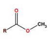
- Fatty acid methyl esters (FAMEs) are easily analyzed by GC, which is undoubtedly the most important analytical method for them. Reliable identification and quantification is readily achieved with MS and flame ionization detectors. However, fatty acids with very long chains and high number of double bonds are thermally unstable, which make their GC analysis impossible. HPLC/MS is then an analytical method of choice.
- FAMEs are separated in NARP systems using C18-type columns.
- APCI, APPI as well as ESI ionize them, but substantial differences in sensitivity exist. Positive ion APCI spectra show protonated molecule [M+H]+ and fragments mostly formed by loss of methanol and further elimination of water. Low mass ions result from the fragmentation of the hydrocarbon chain. APPI provides similar spectra whereas ESI spectra look more complex showing also various adducts with mobile phase components.
Monoacylglycerols (MGs)
Monoacylglycerols (MGs) are simple lipids consisting of one fatty acid covalently bonded to glycerol. They are found in low amounts in most tissues and e.g., 2-MGs are known to be major products of the intestinal digestion of dietary fats. Besides analysis in biological samples they are also monitored in commercial surfactants or samples from biodiesel production. 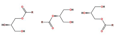
Analysis of MGs is complicated by rapid migration of acyl on the glycerol backbone, which results in a mixture containing all isomers in equilibrium. Therefore, MGs are usually not separated as intact molecules. Instead, total fraction of MGs is isolated, transesterified, and the resulting FAMEs analyzed by GC.
- When analyzed by HPLC/MS, non-aqueous reversed-phase systems with C18 columns and APCI detection are used.
- The base peak in APCI spectra of MGs corresponds to loss of water from the molecular adduct [M+H-H2O]+; other abundant ions are acylium ions [RiCO]+ and acylium ions after loss of water [RiCO-H2O]+.
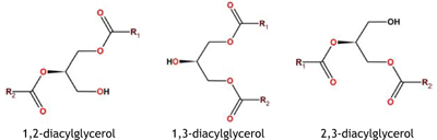
Diacylglycerols (DGs)
Diacylglycerols (DGs) are simple lipids consisting of two fatty acids covalently bonded to glycerol. DGs play important roles in signaling and they are known to be intermediates in enzymatic hydrolysis of TGs. DGs are the main mode of transportation of lipids in insects. Similarly to MGs, spontaneous acyl migration may occur particularly in polar solvents or at elevated temperatures until equilibrium between regioisomers is reached. For this reason, derivatization of free hydroxy group in DGs is recommended. Derivatization is usually achieved by acetylation, but several other derivatization procedures can be found in literature. Derivatization also improves separation or detection properties of DGs.
- Similarly to other acylglycerolipids, NARP systems with C18 columns and APCI, APPI or ESI are used.
- Positive ion mode APCI spectra show abundant [M+H-H2O]+ and [M+H‑RiCOOH]+ ions. Complex lipids, such as phosphoglycerolipids or glycosyldiacylglycerols are sometimes converted to DGs before analysis. Chiral-phase columns are used to separate sn-1,2-diacylglycerols from their sn-2,3 counterparts both directly or after derivatization as various urethanes.
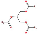
Triacylglycerols (TGs)
Triacylglycerols (TGs) are simple lipids consisting of three fatty acids covalently bonded to glycerol. TGs are perhaps the most frequently analyzed lipids because of their importance for human nutrition and health. They are the main constituents of vegetable oils and animal fats.
- TGs are mostly analyzed in NARP HPLC on octadecyl-modified silica columns.
- The mobile phases usually consist of acetonitrile mixed with stronger solvent (e.g., 2-propanol or acetone) that improves their solubility and affects the selectivity and efficiency of the separation. The retention order generally follows the ECN values. Silver-ion HPLC is used to separate TGs according to the number, geometry and position of the double bonds within FA residues. Mobile phases based on hexane, acetonitrile and 2-propanol or acetone provide good chromatographic resolution.
- Gradient elution is preferred in both chromatographic systems.
- Both APCI and APPI suit well for MS detection. The main fragment ions are diacylglycerol ions [M+H‑RiCOOH]+ originating after loss of a neutral molecule of FA. Besides identification of all FAs bonded to glycerol, information about their regiospecific distribution is obtained from relative intensities of diacylglycerol ions.
- TGs can be also analyzed in HPLC/MS systems with electrospray ionization. Silica columns with C18 chains are used together with mobile phases typically containing methanol and some other organic solvent (e.g., acetonitrile or chloroform). Because ESI spectra do not show protonated molecules, cationization reagents with ammonium (I) or sodium (I) ions are used to induce molecular adduct ions formation. Fragment ions are usually missing or have very low intensities.
Sterols
Sterols are terpenoid lipids – steroids - with a hydroxyl group at the 3-position of the A-ring. They occur ubiquitously in both animals and plants as essential membrane components. The most abundant sterol in animal tissues is cholesterol, which modulates the fluidity of membranes. Certain plant sterols act as growth hormones and have crucial effects on plant development. 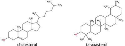
- Sterols are analyzed in reversed-phase systems with C8 or C18 columns.
- Mobile phases typically consist of methanol and water, sometimes containing ammonium acetate to promote formation of ammonium adducts. 2-Propanol can be used for gradient elution.
- All API ionizations (ESI, APCI or APPI) provide mass spectra that mostly show [M+H-H2O]+ as the base peak.
Glycerophospholipids (PLs)
PLs are major components of all biological membranes and play important roles in many biological processes.
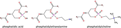
Glycerophospholipids are complex lipids derived mostly from sn-1,2-diacylglycerols, with a phosphate group in the position sn-3 linked to an amino-alcohol, carbohydrate, amino acid or other group. Therefore, several distinct molecular classes of PLs exist. The main PLs are:
- phosphatidylcholines (PCs),
- phosphatidylethanolamines (PEs),
- phosphatidylinositols (PIs),
- phosphatidylserines (PSs),
- phosphatidylglycerols (PGs),
- phosphatidic acids (PAs)
- cardiolipins (CLs).
They are best separated into their respective classes by NP-HPLC using plane silicas or phases chemically modified with diol or amino groups.
- Mobile phases are composed of solvents such as hexane, acetonitrile, chloroform, 2-propanol or methanol, and sometimes contain buffer to adjust pH.
- Gradient elutions are commonly used. For separation of PL species, numerous RP-HPLC systems were developed utilizing silica-based C8, C18 and C30 columns, or non-polar polymeric stationary phases. Mobile phases usually contain methanol and - depending on a particular PL class - other components like water or another organic solvent.
- PLs are preferably ionized by ESI, though problems related to limited compatibility of the mobile phase with the ion source sometimes occur. APCI is used to some extent, but gentle ESI usually provides better spectra. Depending on the PL structures, positive or negative ion mode is preferred. Some PLs are zwitterionic (e.g., PEs) and can be ionized in both modes. Protonated, metalated or ammoniated molecules are commonly generated in the positive ion mode. Alkali metal ions are sometimes intentionally added to the mobile phase to form adducts providing more informative MS/MS spectra than the protonated molecules.
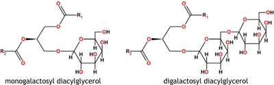
Glyceroglycolipids (GLs)
Galactosyl diacylglycerols together with sulphoquinovosyl diacylglycerols are the most abundant lipids in all photosynthetic tissues. They are components of the membrane systems in chloroplast of plants, algae and some bacteria. Monogalactosyl diacylglycerols (MGDGs) and digalactosyl diacylglycerols (DGDGs) consisting of diacylglycerols connected by a glycosidic bond to a carbohydrate moiety are the most common GLs.
Class separation of plant lipids is challenging, particularly when attempted to separate all of them in a single HPLC run.
- Stationary phases for NP separations are based on plain silica, diol or polyvinyl alcohol functionalized silica materials.
- MGDG and DGDG molecular species are analyzed in RP systems in methanol/water or methanol/acetonitrile/water systems.
- Isocratic elution conditions are sometimes preferred, but gradient methods work equally well.
- GLs are best ionized by ESI providing mostly sodium adducts [M+Na]+. Ammonium salts are sometimes added to the mobile phase generate ammonium adducts for subsequent fragmentations.
There are several good reasons to perform chemical derivatization of lipids before their HPLC/MS analysis. Suitable modification can substantially improve signal intensity and thus increase detection limits. Separation properties of lipids might be also improved and separation of enantiomers is made possible. Derivatization is sometimes used to avoid spontaneous acyl migration, e.g., by acetylation of free hydroxyl group in DGs.
Derivatization is useful for structural elucidation of lipids. Typical examples are derivatives that allow for locating position(s) of double bond(s). Some of the reactions are known for a long time from GC/MS, particularly those for derivatization of FAs. For example, picolinyl esters of very long FAs can be easily separated by RP-HPLC and their APCI spectra provide information about double bond position (s).
Formation of ozonides and vicinal diols is particularly useful in this respect. The reaction with ozone can be performed in the gas phase, directly in the mass spectrometer. Two versions of this approach, ozone ESI MS performed in the ion source and ozone-induced dissociation, taking place in the ion trap analyzer were recently demonstrated.
Once lipids are identified, their quantification might be of interest. Quantification is usually not an easy task, because a large number of lipids is found in samples. Therefore, it is not convenient or even possible to work with standards for all components to perform “classical” quantification. Instead, response factors for individual lipids are used.
The response factors are known to depend on the lipid structure. Nature of polar headgroup, number of double bonds, length of the fatty acid chain or regiospecific distribution of acyls on the glycerol backbone affect them substantially. Response factors were intensively studied in TGs, because their quantification is of interest in many areas, including industrial applications.
APCI response factors of saturated single-acid TGs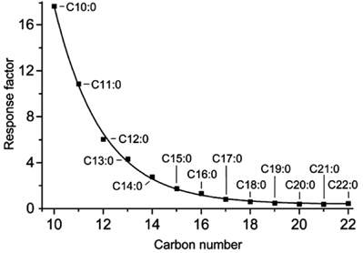 adapted from Holčapek et al., J. Sep. Sci. 2005
Quantification based on peak area without correction by response factors is used when amounts of the same lipids in two samples are directly compared, e.g., when comparing samples from normal and diseased cells. Quantification of one or several lipids (e.g., fytosterols) is done by standard procedures utilizing standards and internal standards. Common procedures based on calibration curves are then used.
MS/MS can be utilized to achieve better limit of detection and get higher specificity.
Alongside with classical analytical procedures for lipids, that are laborious and time consuming, a new approach is being developed. Recent technological advancement of MS allows us analyzing complex lipid samples directly, without any chromatographic separation.
This “shotgun lipidomic” approach utilizes mass spectrometers with electrospray ionization, MS/MS and exact mass measurement capability:
- Total lipid extracts are directly infused; lipids are ionized and subjected to fragmentation.
- Large amounts of data are then interpreted with the aid of sophisticated software tools.
- The
 Lipidomic approach is highly useful, because allows for rapid profiling of hundreds or thousands components at a time.
The scope of this chapter is limited just to a brief introduction into the field of lipid analysis. Readers looking for more information can consult numerous books or websites. Some of them that are good to start with are listed bellow.
- Modern Methods for Lipid Analysis by Liquid Chromatography. William Craig Byrdwell (Editor) AOCS Press; Champaign, Illinois (2005).
- Lipid Analysis - third edition. Christie, W.W. The Oily Press, Bridgwater (2003).
- Advances in Lipid Methodology - One to Four. Christie, W.W. (Editor) The Oily Press, Ayr/Dundee. (1992 to 1997).
- The lipid library. URL: http://www.lipidlibrary.co.uk/
- Cyberlipid center. URL: http://www.cyberlipid.org/

